Evolution of heart and aortic arches in vertebrates pdf
25/03/2018 · As per evolutionary biology Heart is a modified blood vessel. In Pisces the heart is two chambered, in amphibians it is three chambered, whereas in reptiles it is incompletely divided four
problems in the cephalic neural crest may result in problems affecting both pharyngeal arches and heart. Many syndromes have combinations of craniofacial and heart malformations, because of the commonality of their neural crest cells .
Vertebrate Circulation Bi 150/150.1 . Functions Transport Gasses Heat Metabolic products Hormones Wastes Blood . Origin of the Cardiovascular System Mesoderm or mesenchyme gives rise to blood islands and the muscular heart. Blood islands: precursors of blood vessels (angiogenesis) and blood cells (hematopoiesis) Heart: immediately contractile; has four embryonic chambers: sinus venosus, …
The basic fundamental plan of the aortic arches is similar in different vertebrates during embryonic stages. But in adult the condition of the arrangement is changed either being lost or modified considerably. The number of aortic arches is gradually reduced as the scale of evolution of vertebrates …
17/07/2013 · This video should help students get a grasp on the ridiculously complex series of events that take place during development of the large vessels. From the small aortic arches in …
After emerging from the heart, the aortic artery divides into the right and left dorsal branches. Each branch feeds into a set of arches which are unique to the embryo. Most higher vertebrates have have 6 pairs of aortic arches. In the mammal the 5th pair do not form. These arches evolve to form some of the structures of the mammalian circulation. The fate of each arch varies. Arches 1 & 2
The morphology and the function of the heart and cardiovascular systems of vertebrates have undergone large evolutionary changes associated with the transition between water and air-breathing and during the evolution of endothermy within mammals and birds , .
Necturus Heart and Aortic Arches Carefully skin the insides of the pharynx, top and bottom, concentrating on the area between the gills to the transverse septa. Expose the heart on the ventral floor of the mouth and the afferent arteries leading to the gills.
aortic arches In fish, the arteries supplying the gills, passing from the ventral aorta and then uniting to form the dorsal aorta. In tetrapods they are modified and reduced in number. In tetrapods they are modified and reduced in number.
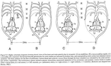
As illustrated in the first figure above (compare hearts C and D of “A detailed view of the heart and aortic arches”), the crocodilian heart is a slight modification of the ancestral reptilian state. So is the mammalian heart, and the bird heart. The bird heart is similar to the crocodilian heart, as Liem and Walker explain: “the ancestors of birds had a heart and pattern of aortic arches
BIOLOGY OF CHORDATES EDUCATIONAL PUBLISHERS

Evolution of the Cardiovascular Autonomic Nervous System
It is hypothesized that the SV is not homologous to that of other vertebrates which could have important implications for understanding heart evolution. In M. glutinosa and E. stoutii, the
At some point during vertebrate evolution from species dwelling in water to living on land, the ancestral double or right aortic arches became single and left-sided in mammals, including humans, as the result of synchronous developments in cardiovascular and respiratory embryogenesis.
The aortic arches (Plates 45, 46, 108–110, 119 Plate 45 Plate 46 Plate 108 Plate 109 Plate 110 Plate 119) are the blood vessels that supply the pharyngeal arches (Chapter 8), and they serve as a communication between the ventral and dorsal aortae.
The cardiac neural crest cells (CNCCs) have played an important role in the evolution and development of the vertebrate cardiovascular system: from reinforcement of the developing aortic arch arteries early in vertebrate evolution, to later orchestration of aortic arch artery remodeling into the great arteries of the heart, and finally outflow
GENETICS AND MOLECULAR BIOLOGY – Gene Expression and Embryogenesis in Amphibians – Horst Grunz Gene Expression and Embryogenesis in Amphibians – Horst Grunz ©Encyclopedia of Life Support Systems (EOLSS) DeRobertis, 1997; Grunz, 1999). Comparatives studies on the function of homologeous genes in invertebrates (Drosophila) and vertebrates (Xenopus) suggest that there …
In both the human embryo and fish the pharynx is bounded by visceral or branchial arches, which are separated by depressions (human embryos) or clefts (fishes) ; in both the heart is situated under the pharynx, and from the ventral aorta, aortic arches pass up on each side, one in each visceral arch, to terminate in the dorsal aortae. In fishes the aortic arches give off vessels to the gills
The structural characteristics of the heart and great arterial vessels amongst living vertebrates do not merely possess surface similarities. Two crucial points need to be emphasized here. First, the retention of “aortic arch arteries” (or “branchial arch arteries”) in non-aquatic vertebrates serves no respiratory function. They are merely connecting pipes. Their sole purpose is to be
The pharyngeal arches and their segmental arrangement are highly conserved throughout evolution from invertebrate chordates such as amphioxus, through to vertebrate agnathans including avians, squamates and mammals. The structural organization of the pharyngeal arches is also highly conserved and involves contributions from each of the three primary endoderm, mesoderm and ectoderm germ …
The left fourth PAA gives rise to the aortic arch while the right PAA gives rise to the subclavian artery and contributes to the pulmonary arteries (light blue). Finally, the sixth PAA gives rise to the ductus arteriosus (dark blue).
ii) Left systemic aorta (Left aortic arch from the left ventricle) Right aortic arch is absent. iii) Left systemic trunk (arise from right side of ventricle) All the trunks are united by connective tissue.
Afferent branchial arteries develop from aortic arches 3, 4, 5, and 6 to supply blood to the gills. The conus arteriosus is a muscular extension of the ventricle which leads into the ventral aorta. At the posterior end of the heart is the sinus venosus, a thin walled space where blood from the veins gathers before entering the atrium.
Circulatory system in vertebrates has two parts, the blood vascular system and lymphatic system, the former includes heart, arteries, veins and blood inside and the latter includes lymph channels and spaces, lymph hearts, lymph nodes and lymph flowing in them from the tissues towards heart. Lymph channels eventually open into larger veins.
Pharyngeal arches L.Moss-Salentijn Pharyngeal arches: a definition A A segmental series of five paired swellings that surround the foregut between days 20 to 35 of embryonic development. These segments, which are unique to vertebrates, are “wedged” between the developing forebrain and heart. Pharyngeal arches a.k.a. visceral or branchial arches Develop (and disappear asDevelop (and
Comparative anatomy of Aortic ArchesAortic arches are paired blood vessels that emerge from the ventricle of the heart which are basically simila…
GALAXYBIOLOGY)IBDP,)TOPIC)6.2) ) 1)) HEART DISSECTION The external structure- The figure given below depicts the evolution of aortic arch in vertebrates.
o Conus arteries in heart to dampen pulsing motion o Myomeres in w shape o Epaxial and hypaxial muscles The Evolution of the Jaw. Splanchnocranium o Modification of brachial gill arches o 1 st arch modified to get large and then jaw to power ventilation across gills o hyomandibular arch is the second arch that was a support structure for the
In this article we will discuss about modification of aortic arches in various vertebrates. Modification in Fishes: Branches from ventral aorta produce aortic arches, …

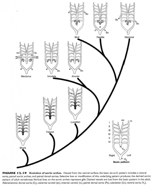
Phylogenetic Overview (12.19) Viewed from the ventral surface, the basic 6-arch pattern includes a ventral aorta, paired aortic arches and paired dorsal aortae. Selective loss or modification of this underlying pattern produces the derived aortic pattern of adult vertebrates.
Structure and functions of Vertebrates Evolution of heart. Evolution of aortic arches and portal systems. Blood circulation in various vertebrate groups. Comparative account of jaw suspensorium and vertebral column. Unit-III- Evolution of urinogenital system. Comparative anatomy of brain. Comparative account of peripheral and autonomous nervous system. Unit-IV- Lateral line system in
The left 4th aortic arch remains as the arcus aortae of the adult while the right one forms the proximal part of the right subclavian artery. 6 aortic arches have always been spoken about. The last two, however, never appear in a prominent arch-shape like the first 4.
Start studying Comparative Anatomy of Vertebrates Chapter 12 Circulatory System. Learn vocabulary, terms, and more with flashcards, games, and other study tools. Learn vocabulary, terms, and more with flashcards, games, and other study tools.
Blood leaves the heart from the ventricle through a single truncus arteriosus which is short and soon branches into two aortic arches which loop left and right and dorsal to the heart to rejoin as a single aorta in the mid dorsal region of the body cavity. Each aortic arch has a branch leading to the lungs and skin where oxygenation occurs. Carotid arteries also branch off the aortic arches
Evolution of Endovascular Aortic Arch Repair Current options for aortic arch repair have grown to encompass several distinct endovascular strategies, including both purely endovascular and “hybrid” endovascular and surgical approaches.
This blog is the fourth piece in a series by Darrel Falk and David Kerk. The previous entry is found here. Let’s start by recalling some aspects of the human heart with which you are probably familiar. The human heart (like that of all mammals) has four pumping chambers – two atria (which
Comparative Anatomy of HeartCirculation Aortic Arches Etc
Aortic Arches in Vertebrates: In various adult vertebrates, the arterial system appears to be different, but they are built on the same basic fundamental plan. The difference is due to increasing complexity of heart due to a change in respiration from gills to lungs.
Evolution of the aortic arches; Evolution of the aortic arches . This diagram shows a side view of the organisms, with the head facing left and heart and lungs to the right. The highly derived patterns of birds and mammals were formed by loss or specialization of the various arches. The order of events leading to each lineage has been reconstructed in some detail. As the caption says, “One of
The heart and the aortic arches are of mesodermal origin. sensory organs: the nasal placodes develop into the olfactory sacs; the audi tory or otic placodes develop into …
In amphibians, the aortic arch is a homologue of the fourth embryonic gill arch innervated by the Xth cranial nerve and the pulmocutaneous artery is a homologue of the sixth embryonic
Comparative anatomy of Aortic Arches Aortic arches are paired blood vessels that emerge from the ventricle of the heart which are basically similar in number and disposition in different vertebrates during the embryonic stages.Kavali.Sc.
The Vertebrate Circulatory System Transportation Respiratory Erythrocytes (red blood cells) transport oxygen from lungs to tissues Hemoglobin of red blood cells is transporter – evolution of the human eye pdf General plan of circulation, evolution of heart and aortic arches Unit 6: Urinogenital System (4) Succession of kidney, Evolution of urinogenital ducts, Types of mammalian uteri.
The role of bilaminar streams in septation of the heart and the importance of hemodynamic factors in the evolution of the aortic arch system have been shown experimentally in the chick embryo. Changes in the hemodynamics of the circulatory system result in cardiovascular malformations.
these vertebrates also have bilaterally symmetrical arch arteries that are remodeled into the asymmetric great arteries of the heart and a singular outflow that becomes the aorta and the pulmonary artery.
Early cardiac development involves the formation of a heart tube, looping of the tube and formation of chambers. These processes are highly similar among all vertebrates, which suggest the existence of evolutionary conservation of the building plan of the heart.
The aortic arches of gill bearing vertebrates are primarily for bringing blood from the heart via the ventral aorta through the gills, where the blood is oxygenated and drained via the efferent branchial arteries into the dorsal aorta for circulation to the body.
Answer and Explanation: 1. (c): Adaptive radiation in evolution was developed by H.F. Osborn in 1898. It is also known as “Divergent evolution” It is development of different functional structures from a common ancestral form.
The role of bilaminar streams in septation of the heart and the importance of hemodynamic factors in the evolution of the aortic arch system have been shown experimentally in the chick embryo
Abstract. Previous studies have shown there to be considerable inter-specific variation in the cardiovascular anatomy of five of the six families of caecilians. Observations on th
The relationship with Evolution of The Heart is in no way to be construed as psychological counseling or any type of psychotherapy. A participant enters into the event and/ or session work with the understanding that he/she is responsible for creating his/or hers own decisions and results.
The authors also discuss the evolution of vertebrate groups from the earliest extinct ancestors to current living vertebrates. The book contains illustrations to clarify various issues as well as
Trends in vertebrate evolution: Circulatory systems. General characteristics of vertebrate circulatory systems The vertebrate heart Development of the heart Heart chambers Comparative anatomy of the heart Fish Lungfish Amphibians Reptiles Birds Mammals. Summary of cardiac evolution Changes to aortic arches Fish Lungfish Anurans Reptiles Birds
Heart size is proportional to body size in all classes of vertebrates except birds where it is proportionally smaller in larger animals. Mammals are the most studied group and the heart …
We discuss these results with regard to the development and evolution of the multichambered vertebrate heart. What did the researchers find? Mesp, which in most vertebrates is involved in cardiac development, is in Ciona limited to a single pair of blastomeres (B7.5).
In fact, the basis for their claims about the aortic arches is most likely to be Michael Denton’s Evolution: A Theory In Crisis (1985, Adler & Adler Publishers: Chevy Chase, MD), which spends several pages arguing the implausibility of evolutionary explanations of heart and aortic arch anatomy.
Aortic arches III, IV, VI persist. The ventral aorta subdivides into three vessels leaving the heart: -pulmonary arch, -the left systemic arch -the right systemic arch. Aortic arch III remains as a component of the carotids. The left systemic arch is composed of the left aortic arch IV and left dorsal aorta. The right systemic arch is composed of the right aortic arch IV and a portion of the
Biology 340 Comparative Embryology Lecture 10 Dr. Stuart Sumida Further Development of the Mesoderm (and Endoderm) Further Development: •Digestive System – Foregut, Midgut, Hindgut •Heart and Aortic Arches •Excretory and Reproductive Systems . Further Development: Digestive System – Foregut, Midgut, Hindgut . The brain grows at an incredible rate. It grows so fast that it makes the
First, the retention of “aortic arch arteries” (or “branchial arch arteries”) in non-aquatic vertebrates serves no respiratory function. They are merely connecting pipes. Their sole purpose is to be used as building blocks to construct modified circulatory elements which function in the species which possess them. But remember, in principle, such “building blocks” might have been
Other arches have been lost and modified through evolution to result in the joining of the ventral and dorsal aorta by the fourth aortic arch, which is now known as the arch of aorta (Holmes, 1975). The lungs of amphibians cannot be relied on for all of their air supply, and other pathways are therefore necessary (Holmes, 1975 ).
Figure 1 As you study the evolution of the aortic arches in vertebrates, observe how these structures of blood supply and drainage became modified as other structures and functions of the animals changed.
Evolution of the Aortic Arch System: Aortic Arches-Fishes/General Model The series of paired vessels that run dorsally through the pharyngeal region; connect the ventral aorta to the dorsal aorta. Each afferent artery typically breaks-up into a capillary bed ventrally at …
Heart Basic Vertebrate Pattern Phylogeny and Evolution of
aortic arches of permanently water-dwelling piscine ancestors, of intermediate forms which occasionally left the water and of primitive tetrapods capable of spending …
The vertebrate heart has undergone an extensive evolutionary remodeling from a piscine cardiac design with a single atrium and a single ventricle to a double cir- culatory system in air-breathing vertebrates with two separate atria and a partial or
In order to gain insights into how the aortic arches changed during the transition of vertebrates to land, transformations of the aortic arches during the metamorphosis of Pelobates fuscus were
determine what led to the evolution of such a heart and high blood pressure; these features could have evolved initially in support of endothermy, large body size, or both.
The blood vessels, capillaries and sinusoids, the heart, development, evolution of aortic arches and heart of vertebrates ; the typical plan and the plan in different vertebrates, comparative anatomy of the heart and arteries, evolution of veins, comparative anatomy
In order to gain insights into how the aortic arches changed during the transition of vertebrates to land, transformations of the aortic arches during the metamorphosis of Pelobates fuscus were investigated and compared with data from the early development of a recent ganoid fish Amia calva and a primitive caudate amphibian Salamandrella
Evolutionary and Developmental Origins of the Cardiac
Circulatory System in Vertebrates (With Diagram
Medical Embryology Development of the Aortic Arches and
Vertebrate Circulation Acads

Comparative Anatomy of Vertebrates Chapter 12 Circulatory


M.Sc. Zoology First Semester Paper-I Paper code Zoo 511
Trends in organ systems Vertebrate circulatory systems
evolution of management information system pdf – (PDF) The vertebrate heart An evolutionary perspective
Vertebrate Physiology Circulation Francis Marion University

Evolution of the Heart
The evolution of amphibian metamorphosis insights
Development and evolution of the pharyngeal apparatus
Peripheral arterial chemoreceptors and the evolution of
The Vertebrate Circulatory System Transportation Respiratory Erythrocytes (red blood cells) transport oxygen from lungs to tissues Hemoglobin of red blood cells is transporter
Evolution of the Aortic Arch System: Aortic Arches-Fishes/General Model The series of paired vessels that run dorsally through the pharyngeal region; connect the ventral aorta to the dorsal aorta. Each afferent artery typically breaks-up into a capillary bed ventrally at …
problems in the cephalic neural crest may result in problems affecting both pharyngeal arches and heart. Many syndromes have combinations of craniofacial and heart malformations, because of the commonality of their neural crest cells .
General plan of circulation, evolution of heart and aortic arches Unit 6: Urinogenital System (4) Succession of kidney, Evolution of urinogenital ducts, Types of mammalian uteri.
The cardiac neural crest cells (CNCCs) have played an important role in the evolution and development of the vertebrate cardiovascular system: from reinforcement of the developing aortic arch arteries early in vertebrate evolution, to later orchestration of aortic arch artery remodeling into the great arteries of the heart, and finally outflow
Comparative anatomy of Aortic Arches Aortic arches are paired blood vessels that emerge from the ventricle of the heart which are basically similar in number and disposition in different vertebrates during the embryonic stages.Kavali.Sc.
aortic arches In fish, the arteries supplying the gills, passing from the ventral aorta and then uniting to form the dorsal aorta. In tetrapods they are modified and reduced in number. In tetrapods they are modified and reduced in number.
In fact, the basis for their claims about the aortic arches is most likely to be Michael Denton’s Evolution: A Theory In Crisis (1985, Adler & Adler Publishers: Chevy Chase, MD), which spends several pages arguing the implausibility of evolutionary explanations of heart and aortic arch anatomy.
Aortic Arches in Vertebrates: In various adult vertebrates, the arterial system appears to be different, but they are built on the same basic fundamental plan. The difference is due to increasing complexity of heart due to a change in respiration from gills to lungs.
Answer and Explanation: 1. (c): Adaptive radiation in evolution was developed by H.F. Osborn in 1898. It is also known as “Divergent evolution” It is development of different functional structures from a common ancestral form.
The aortic arches (Plates 45, 46, 108–110, 119 Plate 45 Plate 46 Plate 108 Plate 109 Plate 110 Plate 119) are the blood vessels that supply the pharyngeal arches (Chapter 8), and they serve as a communication between the ventral and dorsal aortae.
In this article we will discuss about modification of aortic arches in various vertebrates. Modification in Fishes: Branches from ventral aorta produce aortic arches, …
We discuss these results with regard to the development and evolution of the multichambered vertebrate heart. What did the researchers find? Mesp, which in most vertebrates is involved in cardiac development, is in Ciona limited to a single pair of blastomeres (B7.5).
Phylogenetic Overview (12.19) Viewed from the ventral surface, the basic 6-arch pattern includes a ventral aorta, paired aortic arches and paired dorsal aortae. Selective loss or modification of this underlying pattern produces the derived aortic pattern of adult vertebrates.
(PDF) Hemodynamic factors in normal and abnormal
The Evolution of the Heart in Vertebrates o 6 pairs
In fact, the basis for their claims about the aortic arches is most likely to be Michael Denton’s Evolution: A Theory In Crisis (1985, Adler & Adler Publishers: Chevy Chase, MD), which spends several pages arguing the implausibility of evolutionary explanations of heart and aortic arch anatomy.
Necturus Heart and Aortic Arches Carefully skin the insides of the pharynx, top and bottom, concentrating on the area between the gills to the transverse septa. Expose the heart on the ventral floor of the mouth and the afferent arteries leading to the gills.
The blood vessels, capillaries and sinusoids, the heart, development, evolution of aortic arches and heart of vertebrates ; the typical plan and the plan in different vertebrates, comparative anatomy of the heart and arteries, evolution of veins, comparative anatomy
The role of bilaminar streams in septation of the heart and the importance of hemodynamic factors in the evolution of the aortic arch system have been shown experimentally in the chick embryo. Changes in the hemodynamics of the circulatory system result in cardiovascular malformations.
This blog is the fourth piece in a series by Darrel Falk and David Kerk. The previous entry is found here. Let’s start by recalling some aspects of the human heart with which you are probably familiar. The human heart (like that of all mammals) has four pumping chambers – two atria (which
The aortic arches of gill bearing vertebrates are primarily for bringing blood from the heart via the ventral aorta through the gills, where the blood is oxygenated and drained via the efferent branchial arteries into the dorsal aorta for circulation to the body.
Biology 340 Comparative Embryology Lecture 10 Dr. Stuart Sumida Further Development of the Mesoderm (and Endoderm) Further Development: •Digestive System – Foregut, Midgut, Hindgut •Heart and Aortic Arches •Excretory and Reproductive Systems . Further Development: Digestive System – Foregut, Midgut, Hindgut . The brain grows at an incredible rate. It grows so fast that it makes the
At some point during vertebrate evolution from species dwelling in water to living on land, the ancestral double or right aortic arches became single and left-sided in mammals, including humans, as the result of synchronous developments in cardiovascular and respiratory embryogenesis.
these vertebrates also have bilaterally symmetrical arch arteries that are remodeled into the asymmetric great arteries of the heart and a singular outflow that becomes the aorta and the pulmonary artery.
aortic arches of permanently water-dwelling piscine ancestors, of intermediate forms which occasionally left the water and of primitive tetrapods capable of spending …
Comparative anatomy of Aortic Arches Aortic arches are paired blood vessels that emerge from the ventricle of the heart which are basically similar in number and disposition in different vertebrates during the embryonic stages.Kavali.Sc.
Answer and Explanation: 1. (c): Adaptive radiation in evolution was developed by H.F. Osborn in 1898. It is also known as “Divergent evolution” It is development of different functional structures from a common ancestral form.
Other arches have been lost and modified through evolution to result in the joining of the ventral and dorsal aorta by the fourth aortic arch, which is now known as the arch of aorta (Holmes, 1975). The lungs of amphibians cannot be relied on for all of their air supply, and other pathways are therefore necessary (Holmes, 1975 ).
Pharyngeal arches L.Moss-Salentijn Pharyngeal arches: a definition A A segmental series of five paired swellings that surround the foregut between days 20 to 35 of embryonic development. These segments, which are unique to vertebrates, are “wedged” between the developing forebrain and heart. Pharyngeal arches a.k.a. visceral or branchial arches Develop (and disappear asDevelop (and
The Role of Neural Crest Cells in Vertebrate Cardiac
Modification of Aortic Arches in Vertebrates Discussed!
The relationship with Evolution of The Heart is in no way to be construed as psychological counseling or any type of psychotherapy. A participant enters into the event and/ or session work with the understanding that he/she is responsible for creating his/or hers own decisions and results.
The left fourth PAA gives rise to the aortic arch while the right PAA gives rise to the subclavian artery and contributes to the pulmonary arteries (light blue). Finally, the sixth PAA gives rise to the ductus arteriosus (dark blue).
Early cardiac development involves the formation of a heart tube, looping of the tube and formation of chambers. These processes are highly similar among all vertebrates, which suggest the existence of evolutionary conservation of the building plan of the heart.
The role of bilaminar streams in septation of the heart and the importance of hemodynamic factors in the evolution of the aortic arch system have been shown experimentally in the chick embryo
The structural characteristics of the heart and great arterial vessels amongst living vertebrates do not merely possess surface similarities. Two crucial points need to be emphasized here. First, the retention of “aortic arch arteries” (or “branchial arch arteries”) in non-aquatic vertebrates serves no respiratory function. They are merely connecting pipes. Their sole purpose is to be
aortic arches of permanently water-dwelling piscine ancestors, of intermediate forms which occasionally left the water and of primitive tetrapods capable of spending …
Trends in vertebrate evolution Circulatory systems
The Galaxy School biology4isc.weebly.com
The aortic arches (Plates 45, 46, 108–110, 119 Plate 45 Plate 46 Plate 108 Plate 109 Plate 110 Plate 119) are the blood vessels that supply the pharyngeal arches (Chapter 8), and they serve as a communication between the ventral and dorsal aortae.
problems in the cephalic neural crest may result in problems affecting both pharyngeal arches and heart. Many syndromes have combinations of craniofacial and heart malformations, because of the commonality of their neural crest cells .
The authors also discuss the evolution of vertebrate groups from the earliest extinct ancestors to current living vertebrates. The book contains illustrations to clarify various issues as well as
The relationship with Evolution of The Heart is in no way to be construed as psychological counseling or any type of psychotherapy. A participant enters into the event and/ or session work with the understanding that he/she is responsible for creating his/or hers own decisions and results.
Aortic arches III, IV, VI persist. The ventral aorta subdivides into three vessels leaving the heart: -pulmonary arch, -the left systemic arch -the right systemic arch. Aortic arch III remains as a component of the carotids. The left systemic arch is composed of the left aortic arch IV and left dorsal aorta. The right systemic arch is composed of the right aortic arch IV and a portion of the
The Vertebrate Circulatory System Transportation Respiratory Erythrocytes (red blood cells) transport oxygen from lungs to tissues Hemoglobin of red blood cells is transporter
We discuss these results with regard to the development and evolution of the multichambered vertebrate heart. What did the researchers find? Mesp, which in most vertebrates is involved in cardiac development, is in Ciona limited to a single pair of blastomeres (B7.5).
Biology 340 Comparative Embryology Lecture 10 Dr. Stuart Sumida Further Development of the Mesoderm (and Endoderm) Further Development: •Digestive System – Foregut, Midgut, Hindgut •Heart and Aortic Arches •Excretory and Reproductive Systems . Further Development: Digestive System – Foregut, Midgut, Hindgut . The brain grows at an incredible rate. It grows so fast that it makes the
Hemodynamic factors in normal and abnormal cardiovascular
Modification of Aortic Arches in Various Vertebrates Zoology
The structural characteristics of the heart and great arterial vessels amongst living vertebrates do not merely possess surface similarities. Two crucial points need to be emphasized here. First, the retention of “aortic arch arteries” (or “branchial arch arteries”) in non-aquatic vertebrates serves no respiratory function. They are merely connecting pipes. Their sole purpose is to be
First, the retention of “aortic arch arteries” (or “branchial arch arteries”) in non-aquatic vertebrates serves no respiratory function. They are merely connecting pipes. Their sole purpose is to be used as building blocks to construct modified circulatory elements which function in the species which possess them. But remember, in principle, such “building blocks” might have been
The basic fundamental plan of the aortic arches is similar in different vertebrates during embryonic stages. But in adult the condition of the arrangement is changed either being lost or modified considerably. The number of aortic arches is gradually reduced as the scale of evolution of vertebrates …
Structure and functions of Vertebrates Evolution of heart. Evolution of aortic arches and portal systems. Blood circulation in various vertebrate groups. Comparative account of jaw suspensorium and vertebral column. Unit-III- Evolution of urinogenital system. Comparative anatomy of brain. Comparative account of peripheral and autonomous nervous system. Unit-IV- Lateral line system in
70 responses to “Evolution of heart and aortic arches in vertebrates pdf”
As illustrated in the first figure above (compare hearts C and D of “A detailed view of the heart and aortic arches”), the crocodilian heart is a slight modification of the ancestral reptilian state. So is the mammalian heart, and the bird heart. The bird heart is similar to the crocodilian heart, as Liem and Walker explain: “the ancestors of birds had a heart and pattern of aortic arches
heart and aortic arches of rhinatrematid caecilians
Circulatory system in vertebrates has two parts, the blood vascular system and lymphatic system, the former includes heart, arteries, veins and blood inside and the latter includes lymph channels and spaces, lymph hearts, lymph nodes and lymph flowing in them from the tissues towards heart. Lymph channels eventually open into larger veins.
Aortic Arches Anatomy & Physiology – WikiVet English
GENETICS AND MOLECULAR BIOLOGY – Gene Expression and Embryogenesis in Amphibians – Horst Grunz Gene Expression and Embryogenesis in Amphibians – Horst Grunz ©Encyclopedia of Life Support Systems (EOLSS) DeRobertis, 1997; Grunz, 1999). Comparatives studies on the function of homologeous genes in invertebrates (Drosophila) and vertebrates (Xenopus) suggest that there …
The vertebrate heart an evolutionary perspective
The Galaxy School biology4isc.weebly.com
LAB 8 CIRCULATORY AND RESPIRATORY SYSTEMS
Trends in vertebrate evolution: Circulatory systems. General characteristics of vertebrate circulatory systems The vertebrate heart Development of the heart Heart chambers Comparative anatomy of the heart Fish Lungfish Amphibians Reptiles Birds Mammals. Summary of cardiac evolution Changes to aortic arches Fish Lungfish Anurans Reptiles Birds
The left-sided aortic arch in humans viewed as the end
The evolution of amphibian metamorphosis Insights based
LAB 8 CIRCULATORY AND RESPIRATORY SYSTEMS
The vertebrate heart has undergone an extensive evolutionary remodeling from a piscine cardiac design with a single atrium and a single ventricle to a double cir- culatory system in air-breathing vertebrates with two separate atria and a partial or
Pharyngeal arches a definition Pharyngeal arches A
these vertebrates also have bilaterally symmetrical arch arteries that are remodeled into the asymmetric great arteries of the heart and a singular outflow that becomes the aorta and the pulmonary artery.
The Vertebrate Circulatory System
(PDF) Hemodynamic factors in normal and abnormal
Evolution of the aortic arches NCSE
In both the human embryo and fish the pharynx is bounded by visceral or branchial arches, which are separated by depressions (human embryos) or clefts (fishes) ; in both the heart is situated under the pharynx, and from the ventral aorta, aortic arches pass up on each side, one in each visceral arch, to terminate in the dorsal aortae. In fishes the aortic arches give off vessels to the gills
Comparative Anatomy of HeartCirculation Aortic Arches Etc
Peripheral arterial chemoreceptors and the evolution of
Evidences for Evolution Part 3a The Heart and
Structure and functions of Vertebrates Evolution of heart. Evolution of aortic arches and portal systems. Blood circulation in various vertebrate groups. Comparative account of jaw suspensorium and vertebral column. Unit-III- Evolution of urinogenital system. Comparative anatomy of brain. Comparative account of peripheral and autonomous nervous system. Unit-IV- Lateral line system in
Aortic arches Embryology
o Conus arteries in heart to dampen pulsing motion o Myomeres in w shape o Epaxial and hypaxial muscles The Evolution of the Jaw. Splanchnocranium o Modification of brachial gill arches o 1 st arch modified to get large and then jaw to power ventilation across gills o hyomandibular arch is the second arch that was a support structure for the
(PDF) The vertebrate heart An evolutionary perspective
aortic arches of permanently water-dwelling piscine ancestors, of intermediate forms which occasionally left the water and of primitive tetrapods capable of spending …
Modification of Aortic Arches in Various Vertebrates Zoology
The evolution of amphibian metamorphosis Insights based
Phylogenetic Overview (12.19) Viewed from the ventral surface, the basic 6-arch pattern includes a ventral aorta, paired aortic arches and paired dorsal aortae. Selective loss or modification of this underlying pattern produces the derived aortic pattern of adult vertebrates.
Evolution of the Heart
Aortic Arches an overview ScienceDirect Topics
Aortic Arches Encyclopedia.com
The cardiac neural crest cells (CNCCs) have played an important role in the evolution and development of the vertebrate cardiovascular system: from reinforcement of the developing aortic arch arteries early in vertebrate evolution, to later orchestration of aortic arch artery remodeling into the great arteries of the heart, and finally outflow
Evidences for Evolution Part 3a The Heart and
The evolution of amphibian metamorphosis Insights based
Circulatory System in Vertebrates (With Diagram
17/07/2013 · This video should help students get a grasp on the ridiculously complex series of events that take place during development of the large vessels. From the small aortic arches in …
(PDF) The vertebrate heart An evolutionary perspective
(PDF) Hemodynamic factors in normal and abnormal
Hearts Home NCSE
First, the retention of “aortic arch arteries” (or “branchial arch arteries”) in non-aquatic vertebrates serves no respiratory function. They are merely connecting pipes. Their sole purpose is to be used as building blocks to construct modified circulatory elements which function in the species which possess them. But remember, in principle, such “building blocks” might have been
Modification of Aortic Arches in Vertebrates Discussed!
Heart Basic Vertebrate Pattern Phylogeny and Evolution of
Comparative Review The Phylotypic Stage of Vertebrates
The aortic arches of gill bearing vertebrates are primarily for bringing blood from the heart via the ventral aorta through the gills, where the blood is oxygenated and drained via the efferent branchial arteries into the dorsal aorta for circulation to the body.
Development and evolution of the pharyngeal apparatus
The Functional Signi Þ cance of the Reptilian Heart New
The Evolution of the Heart in Vertebrates o 6 pairs
25/03/2018 · As per evolutionary biology Heart is a modified blood vessel. In Pisces the heart is two chambered, in amphibians it is three chambered, whereas in reptiles it is incompletely divided four
(PDF) Hemodynamic factors in normal and abnormal
Circulatory System in Vertebrates (With Diagram
The left-sided aortic arch in humans viewed as the end
At some point during vertebrate evolution from species dwelling in water to living on land, the ancestral double or right aortic arches became single and left-sided in mammals, including humans, as the result of synchronous developments in cardiovascular and respiratory embryogenesis.
Evolution of the aortic arches NCSE
Evolutionary and Developmental Origins of the Cardiac
Trends in vertebrate evolution Circulatory systems
Start studying Comparative Anatomy of Vertebrates Chapter 12 Circulatory System. Learn vocabulary, terms, and more with flashcards, games, and other study tools. Learn vocabulary, terms, and more with flashcards, games, and other study tools.
Evolution of the aortic arches NCSE
Comparative Anatomy of Aortic Arches id.scribd.com
17/07/2013 · This video should help students get a grasp on the ridiculously complex series of events that take place during development of the large vessels. From the small aortic arches in …
Aortic Arches Anatomy & Physiology – WikiVet English
Trends in organ systems Vertebrate circulatory systems
The Evolution of the Heart in Vertebrates o 6 pairs
GENETICS AND MOLECULAR BIOLOGY – Gene Expression and Embryogenesis in Amphibians – Horst Grunz Gene Expression and Embryogenesis in Amphibians – Horst Grunz ©Encyclopedia of Life Support Systems (EOLSS) DeRobertis, 1997; Grunz, 1999). Comparatives studies on the function of homologeous genes in invertebrates (Drosophila) and vertebrates (Xenopus) suggest that there …
Trends in organ systems Vertebrate circulatory systems
The Galaxy School biology4isc.weebly.com
Early cardiac development involves the formation of a heart tube, looping of the tube and formation of chambers. These processes are highly similar among all vertebrates, which suggest the existence of evolutionary conservation of the building plan of the heart.
Evolutionary and Developmental Origins of the Cardiac
Arteries’and’Veins’ USD Biology
Evidence for Endothermic Ancestors of Crocodiles at the
Comparative anatomy of Aortic Arches Aortic arches are paired blood vessels that emerge from the ventricle of the heart which are basically similar in number and disposition in different vertebrates during the embryonic stages.Kavali.Sc.
Modification of Aortic Arches in Various Vertebrates Zoology
Evolution of Endovascular Aortic Arch Repair Current options for aortic arch repair have grown to encompass several distinct endovascular strategies, including both purely endovascular and “hybrid” endovascular and surgical approaches.
M.Sc. Zoology First Semester Paper-I Paper code Zoo 511
Comparative Anatomy of Aortic Arches id.scribd.com
Evidences for Evolution Part 3a The Heart and
Start studying Comparative Anatomy of Vertebrates Chapter 12 Circulatory System. Learn vocabulary, terms, and more with flashcards, games, and other study tools. Learn vocabulary, terms, and more with flashcards, games, and other study tools.
Evolution of Heart in vertebrates YouTube
The evolution of amphibian metamorphosis insights based
aortic arches of permanently water-dwelling piscine ancestors, of intermediate forms which occasionally left the water and of primitive tetrapods capable of spending …
Human Embryology and Morphology 17 Embryology
The Role of Neural Crest Cells in Vertebrate Cardiac
Aortic Arches Encyclopedia.com
Aortic Arches in Vertebrates: In various adult vertebrates, the arterial system appears to be different, but they are built on the same basic fundamental plan. The difference is due to increasing complexity of heart due to a change in respiration from gills to lungs.
Aortic arches Embryology
Heart Basic Vertebrate Pattern Phylogeny and Evolution of
Evolution of the aortic arches NCSE
GENETICS AND MOLECULAR BIOLOGY – Gene Expression and Embryogenesis in Amphibians – Horst Grunz Gene Expression and Embryogenesis in Amphibians – Horst Grunz ©Encyclopedia of Life Support Systems (EOLSS) DeRobertis, 1997; Grunz, 1999). Comparatives studies on the function of homologeous genes in invertebrates (Drosophila) and vertebrates (Xenopus) suggest that there …
Comparative Anatomy of Aortic Arches id.scribd.com
Peripheral arterial chemoreceptors and the evolution of
Phylogenetic Overview (12.19) Viewed from the ventral surface, the basic 6-arch pattern includes a ventral aorta, paired aortic arches and paired dorsal aortae. Selective loss or modification of this underlying pattern produces the derived aortic pattern of adult vertebrates.
The left-sided aortic arch in humans viewed as the end
Evolution of the Cardiovascular Autonomic Nervous System
The evolution of amphibian metamorphosis insights based
Aortic Arches in Vertebrates: In various adult vertebrates, the arterial system appears to be different, but they are built on the same basic fundamental plan. The difference is due to increasing complexity of heart due to a change in respiration from gills to lungs.
Evolution of Heart in vertebrates YouTube
heart and aortic arches of rhinatrematid caecilians
Figure 1 As you study the evolution of the aortic arches in vertebrates, observe how these structures of blood supply and drainage became modified as other structures and functions of the animals changed.
Evolution of the Cardiovascular Autonomic Nervous System
The evolution of amphibian metamorphosis insights
Gene Expression and Embryogenesis in Amphibians EOLSS
Aortic Arches in Vertebrates: In various adult vertebrates, the arterial system appears to be different, but they are built on the same basic fundamental plan. The difference is due to increasing complexity of heart due to a change in respiration from gills to lungs.
Comparative Anatomy of Aortic Arches id.scribd.com
Hemodynamic factors in normal and abnormal cardiovascular
In this article we will discuss about modification of aortic arches in various vertebrates. Modification in Fishes: Branches from ventral aorta produce aortic arches, …
The Vertebrate Circulatory System
The Functional Signi Þ cance of the Reptilian Heart New
Aortic Arches an overview ScienceDirect Topics
The structural characteristics of the heart and great arterial vessels amongst living vertebrates do not merely possess surface similarities. Two crucial points need to be emphasized here. First, the retention of “aortic arch arteries” (or “branchial arch arteries”) in non-aquatic vertebrates serves no respiratory function. They are merely connecting pipes. Their sole purpose is to be
Modification of Aortic Arches in Vertebrates Discussed!
heart and aortic arches of rhinatrematid caecilians
General plan of circulation, evolution of heart and aortic arches Unit 6: Urinogenital System (4) Succession of kidney, Evolution of urinogenital ducts, Types of mammalian uteri.
startpage University of the Cumberlands
The role of bilaminar streams in septation of the heart and the importance of hemodynamic factors in the evolution of the aortic arch system have been shown experimentally in the chick embryo. Changes in the hemodynamics of the circulatory system result in cardiovascular malformations.
The Vertebrate Circulatory System
Comparative Anatomy of Vertebrates Chapter 12 Circulatory
Medical Embryology Development of the Aortic Arches and
This blog is the fourth piece in a series by Darrel Falk and David Kerk. The previous entry is found here. Let’s start by recalling some aspects of the human heart with which you are probably familiar. The human heart (like that of all mammals) has four pumping chambers – two atria (which
Comparative Anatomy of Vertebrates Chapter 12 Circulatory
(PDF) Hemodynamic factors in normal and abnormal
Figure 1 As you study the evolution of the aortic arches in vertebrates, observe how these structures of blood supply and drainage became modified as other structures and functions of the animals changed.
The vertebrate heart an evolutionary perspective
General plan of circulation, evolution of heart and aortic arches Unit 6: Urinogenital System (4) Succession of kidney, Evolution of urinogenital ducts, Types of mammalian uteri.
Circulatory System Questions and Study Guide Quizlet
The Role of Neural Crest Cells in Vertebrate Cardiac
The Galaxy School biology4isc.weebly.com
The pharyngeal arches and their segmental arrangement are highly conserved throughout evolution from invertebrate chordates such as amphioxus, through to vertebrate agnathans including avians, squamates and mammals. The structural organization of the pharyngeal arches is also highly conserved and involves contributions from each of the three primary endoderm, mesoderm and ectoderm germ …
Peripheral arterial chemoreceptors and the evolution of
Vertebrate Physiology Circulation Francis Marion University
Evolution of Heart in vertebrates YouTube
ii) Left systemic aorta (Left aortic arch from the left ventricle) Right aortic arch is absent. iii) Left systemic trunk (arise from right side of ventricle) All the trunks are united by connective tissue.
Evolution of Aortic Arch Repair Europe PMC Article
Circulatory System Questions and Study Guide Quizlet
Vertebrate Physiology Circulation Francis Marion University
these vertebrates also have bilaterally symmetrical arch arteries that are remodeled into the asymmetric great arteries of the heart and a singular outflow that becomes the aorta and the pulmonary artery.
The Functional Signi Þ cance of the Reptilian Heart New
Biology 340 Comparative Embryology Lecture 10 Dr. Stuart
Circulatory system Zoology for IAS IFoS and other
The left 4th aortic arch remains as the arcus aortae of the adult while the right one forms the proximal part of the right subclavian artery. 6 aortic arches have always been spoken about. The last two, however, never appear in a prominent arch-shape like the first 4.
Evolution of Heart in vertebrates YouTube
The authors also discuss the evolution of vertebrate groups from the earliest extinct ancestors to current living vertebrates. The book contains illustrations to clarify various issues as well as
Evidence for Endothermic Ancestors of Crocodiles at the
Gene Expression and Embryogenesis in Amphibians EOLSS
problems in the cephalic neural crest may result in problems affecting both pharyngeal arches and heart. Many syndromes have combinations of craniofacial and heart malformations, because of the commonality of their neural crest cells .
Trends in organ systems Vertebrate circulatory systems
GENETICS AND MOLECULAR BIOLOGY – Gene Expression and Embryogenesis in Amphibians – Horst Grunz Gene Expression and Embryogenesis in Amphibians – Horst Grunz ©Encyclopedia of Life Support Systems (EOLSS) DeRobertis, 1997; Grunz, 1999). Comparatives studies on the function of homologeous genes in invertebrates (Drosophila) and vertebrates (Xenopus) suggest that there …
Evolution of Heart in vertebrates YouTube
Evolution of Endovascular Aortic Arch Repair Current options for aortic arch repair have grown to encompass several distinct endovascular strategies, including both purely endovascular and “hybrid” endovascular and surgical approaches.
The Vertebrate Circulatory System
Trends in organ systems Vertebrate circulatory systems
Modification of Aortic Arches in Vertebrates Discussed!
GENETICS AND MOLECULAR BIOLOGY – Gene Expression and Embryogenesis in Amphibians – Horst Grunz Gene Expression and Embryogenesis in Amphibians – Horst Grunz ©Encyclopedia of Life Support Systems (EOLSS) DeRobertis, 1997; Grunz, 1999). Comparatives studies on the function of homologeous genes in invertebrates (Drosophila) and vertebrates (Xenopus) suggest that there …
The Functional Signi Þ cance of the Reptilian Heart New
Modification of Aortic Arches in Vertebrates Discussed!
Circulatory system Zoology for IAS IFoS and other
This blog is the fourth piece in a series by Darrel Falk and David Kerk. The previous entry is found here. Let’s start by recalling some aspects of the human heart with which you are probably familiar. The human heart (like that of all mammals) has four pumping chambers – two atria (which
startpage University of the Cumberlands
Pharyngeal arches a definition Pharyngeal arches A
Trends in organ systems Vertebrate circulatory systems
The blood vessels, capillaries and sinusoids, the heart, development, evolution of aortic arches and heart of vertebrates ; the typical plan and the plan in different vertebrates, comparative anatomy of the heart and arteries, evolution of veins, comparative anatomy
The evolution of amphibian metamorphosis Insights based
Evolutionary and Developmental Origins of the Cardiac
The aortic arches (Plates 45, 46, 108–110, 119 Plate 45 Plate 46 Plate 108 Plate 109 Plate 110 Plate 119) are the blood vessels that supply the pharyngeal arches (Chapter 8), and they serve as a communication between the ventral and dorsal aortae.
Comparative Anatomy of Aortic Arches id.scribd.com
aortic arches of permanently water-dwelling piscine ancestors, of intermediate forms which occasionally left the water and of primitive tetrapods capable of spending …
Development and evolution of the pharyngeal apparatus
Human Embryology and Morphology 17 Embryology
Biology 340 Comparative Embryology Lecture 10 Dr. Stuart
25/03/2018 · As per evolutionary biology Heart is a modified blood vessel. In Pisces the heart is two chambered, in amphibians it is three chambered, whereas in reptiles it is incompletely divided four
Comparative Anatomy of Vertebrates Chapter 12 Circulatory
The vertebrate heart an evolutionary perspective
Evidences for Evolution Part 3a The Heart and
The relationship with Evolution of The Heart is in no way to be construed as psychological counseling or any type of psychotherapy. A participant enters into the event and/ or session work with the understanding that he/she is responsible for creating his/or hers own decisions and results.
Medical Embryology Development of the Aortic Arches and
In both the human embryo and fish the pharynx is bounded by visceral or branchial arches, which are separated by depressions (human embryos) or clefts (fishes) ; in both the heart is situated under the pharynx, and from the ventral aorta, aortic arches pass up on each side, one in each visceral arch, to terminate in the dorsal aortae. In fishes the aortic arches give off vessels to the gills
The Vertebrate Circulatory System
Modification of Aortic Arches in Various Vertebrates Zoology
The authors also discuss the evolution of vertebrate groups from the earliest extinct ancestors to current living vertebrates. The book contains illustrations to clarify various issues as well as
The left-sided aortic arch in humans viewed as the end
After emerging from the heart, the aortic artery divides into the right and left dorsal branches. Each branch feeds into a set of arches which are unique to the embryo. Most higher vertebrates have have 6 pairs of aortic arches. In the mammal the 5th pair do not form. These arches evolve to form some of the structures of the mammalian circulation. The fate of each arch varies. Arches 1 & 2
Heart Basic Vertebrate Pattern Phylogeny and Evolution of
Evolutionary and Developmental Origins of the Cardiac
We discuss these results with regard to the development and evolution of the multichambered vertebrate heart. What did the researchers find? Mesp, which in most vertebrates is involved in cardiac development, is in Ciona limited to a single pair of blastomeres (B7.5).
Trends in organ systems Vertebrate circulatory systems
Gene Expression and Embryogenesis in Amphibians EOLSS
Vertebrate Circulation Bi 150/150.1 . Functions Transport Gasses Heat Metabolic products Hormones Wastes Blood . Origin of the Cardiovascular System Mesoderm or mesenchyme gives rise to blood islands and the muscular heart. Blood islands: precursors of blood vessels (angiogenesis) and blood cells (hematopoiesis) Heart: immediately contractile; has four embryonic chambers: sinus venosus, …
Evidence for Endothermic Ancestors of Crocodiles at the
In order to gain insights into how the aortic arches changed during the transition of vertebrates to land, transformations of the aortic arches during the metamorphosis of Pelobates fuscus were investigated and compared with data from the early development of a recent ganoid fish Amia calva and a primitive caudate amphibian Salamandrella
Aortic Arches an overview ScienceDirect Topics
Human Embryology and Morphology 17 Embryology
Circulatory system in vertebrates has two parts, the blood vascular system and lymphatic system, the former includes heart, arteries, veins and blood inside and the latter includes lymph channels and spaces, lymph hearts, lymph nodes and lymph flowing in them from the tissues towards heart. Lymph channels eventually open into larger veins.
Evolution of Heart in vertebrates YouTube
Evolution of the Heart
The aortic arches of gill bearing vertebrates are primarily for bringing blood from the heart via the ventral aorta through the gills, where the blood is oxygenated and drained via the efferent branchial arteries into the dorsal aorta for circulation to the body.
Biology 340 Comparative Embryology Lecture 10 Dr. Stuart
Hearts Home NCSE
Blood leaves the heart from the ventricle through a single truncus arteriosus which is short and soon branches into two aortic arches which loop left and right and dorsal to the heart to rejoin as a single aorta in the mid dorsal region of the body cavity. Each aortic arch has a branch leading to the lungs and skin where oxygenation occurs. Carotid arteries also branch off the aortic arches
Evolution of the Cardiovascular Autonomic Nervous System
Development and evolution of the pharyngeal apparatus
Modification of Aortic Arches in Various Vertebrates Zoology
The role of bilaminar streams in septation of the heart and the importance of hemodynamic factors in the evolution of the aortic arch system have been shown experimentally in the chick embryo. Changes in the hemodynamics of the circulatory system result in cardiovascular malformations.
Hemodynamic factors in normal and abnormal cardiovascular
Circulatory system Zoology for IAS IFoS and other
(PDF) Hemodynamic factors in normal and abnormal
The role of bilaminar streams in septation of the heart and the importance of hemodynamic factors in the evolution of the aortic arch system have been shown experimentally in the chick embryo
Human Embryology and Morphology 17 Embryology
Heart size is proportional to body size in all classes of vertebrates except birds where it is proportionally smaller in larger animals. Mammals are the most studied group and the heart …
Aortic Arches Anatomy & Physiology – WikiVet English
The relationship with Evolution of The Heart is in no way to be construed as psychological counseling or any type of psychotherapy. A participant enters into the event and/ or session work with the understanding that he/she is responsible for creating his/or hers own decisions and results.
Evidence for Endothermic Ancestors of Crocodiles at the
heart and aortic arches of rhinatrematid caecilians
This blog is the fourth piece in a series by Darrel Falk and David Kerk. The previous entry is found here. Let’s start by recalling some aspects of the human heart with which you are probably familiar. The human heart (like that of all mammals) has four pumping chambers – two atria (which
Vertebrate Physiology Circulation Francis Marion University
Arteries’and’Veins’ USD Biology
In both the human embryo and fish the pharynx is bounded by visceral or branchial arches, which are separated by depressions (human embryos) or clefts (fishes) ; in both the heart is situated under the pharynx, and from the ventral aorta, aortic arches pass up on each side, one in each visceral arch, to terminate in the dorsal aortae. In fishes the aortic arches give off vessels to the gills
The Evolution of the Heart in Vertebrates o 6 pairs
Evolution of the Heart
these vertebrates also have bilaterally symmetrical arch arteries that are remodeled into the asymmetric great arteries of the heart and a singular outflow that becomes the aorta and the pulmonary artery.
The Functional Signi Þ cance of the Reptilian Heart New
Evolution of the Heart
In order to gain insights into how the aortic arches changed during the transition of vertebrates to land, transformations of the aortic arches during the metamorphosis of Pelobates fuscus were
Biology 340 Comparative Embryology Lecture 10 Dr. Stuart
Medical Embryology Development of the Aortic Arches and